MCL tear surgery in Burr Ridge
Also serving Plainfield, Morris and surrounding areas
What is MCL?

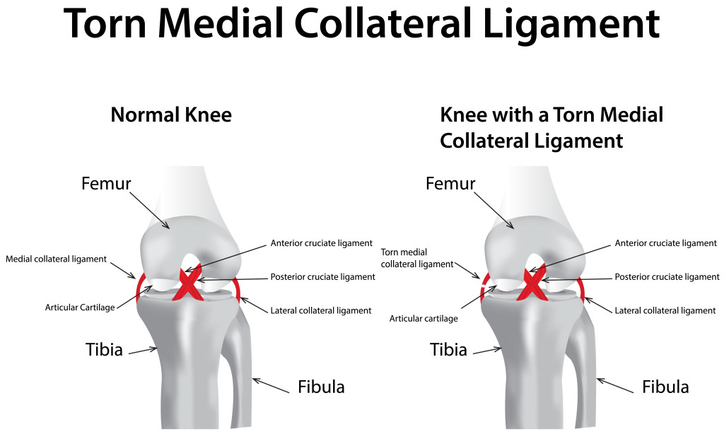
The medial collateral ligament (MCL) is a ligament positioned on the inside of the knee. MCL injuries commonly occur from a blow to the outside of the knee while participating in sports. Pain and swelling may be experienced on the inside of the knee, and some patients will feel some instability to the knee when walking. The majority of MCL injuries are sprains and do not require surgery.
MCL Treatment Options
Non-Surgical Treatment
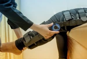
If the MCL injury does not require surgery, then it will be necessary to brace the knee to keep it stable, and attend physical therapy sessions.
Surgical Treatment
Surgery is rarely required for MCL injuries. However, in some cases, a severe injury may actually cause the ligament to pull away from the bone, in which case surgical repair may be required. This injury is commonly seen in football, soccer, and other contact sports. MCL repair surgery entails suturing of the ligament and fixation to bone either above or below the knee with screws or suture anchors. The majority of these injuries are lower grade I or II injuries that heal nicely on their own. Some higher-grade III injuries may require surgery and are often combined ACL/MCL or ACL/PCL/MCL injuries whereby several ligaments will require repair or reconstruction.

MCL Tear Recovery

The non-surgical recovery time of an MCL injury is typically 6-8 weeks with bracing and physical therapy. Surgical recovery and healing will typically take 2-3 months, depending on the severity of injury. Bracing will often be a crucial factor in healing with or without surgery.
Most patients, whether treated surgically or non-surgically, will be able to recover useful function and return to competitive play.
What Are the Types of MCL Tears?
Our board-certified orthopedic surgeon will describe your MCL tear in grades, which are broken into three categories:
- Grade I MCL Tear: A Grade I tear is a mild knee injury that results in a tear of less than 10% of the MCL ligament fibers in the MCL. The knee remains stable with regular movement. Patients with Grade I MCL tear may experience mild pain and tenderness.
- Grade II MCL Tear: A Grade II tear is considered a moderate knee injury where the MCL partially tears. The tear often occurs in the superficial section of the medial collateral ligament. Patients with Grade II MCL tear may feel the knee is loose and lacking stability with movement and experience severe pain and tenderness on the inner side of the knee.
- Grade III MCL Tear: A Grade III tear is a severe knee injury that tears the MCL entirely in both the superficial and deep sections of the ligament. Patients with Grade III MCL tear will have an unstable and loose knee accompanied by intense pain and tenderness. These injuries often coincide with other knee injuries, especially ACL tears.
Dr. Burt will discuss the severity of your MCL tear during your appointment at Midwest Sports Medicine Institute and develop a treatment plan based on his findings.
What Are The Symptoms of a MCL Tear?

Symptoms of a MCL tear vary based on the severity of the knee injury and may include the following:
- Patients may hear a popping sound when the knee injury occurs, whether during a sporting activity or an accident.
- Knee pain develops at the time of the incident or shortly after for most patients.
- The knee may swell and feel stiff with movement after the injury.
- Many patients experience sensations of the knee “giving out” when they put weight or stress on the knee.
- The knee joint may feel like it locks or “catches” when used after an injury.
It’s best to see a healthcare professional immediately after a knee injury to evaluate the damage, determine severity, reduce swelling and create an effective treatment plan that reduces pain and other symptoms.
How Is a MCL Tear Diagnosed?
Dr. Burt can diagnose MCL tear and determine the extent of damage to the ligament. A physical exam and some imaging tests are often necessary to analyze the extent of knee injury and develop a treatment plan.
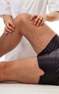
- Physical Examination: Dr. Burt may bend the knee and apply pressure to see if the knee is unstable, which indicates a MCL tear. Palpation on the inner portion of the knee tends to cause pain with MCL tear.
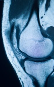
- Imaging Tests: A MRI, ultrasound and/or X-ray is often necessary to diagnose a MCL tear and see if there are other joint structures damaged. Magnetic resonance imaging (MRI) tests are optimal because they detect change in the soft tissue of the knee. Ultrasound and X-ray imaging can detect bone fracture and other injuries.
Who Is More Likely to Experience a MCL Tear?

MCL tears are common among athletes, including skiers, soccer players and football players. While these injuries may not be preventable, exercises that focus on balancing and strengthening the hip and thigh muscles may lower the risk of a MCL tear. Specifically, football linemen may protect themselves from MCL tear by wearing designated braces.
- A MCL tear is likely to happen if one foot is planted on the ground and the lower extremity forcefully shifts direction.
- An object or person hitting the outer side of the knee may tear the MCL at the inside of the knee, such as a football tackle.
- Squatting or lifting heavy objects may cause damage to the MCL.
- Skiing injuries may result in MCL tear by hyperextending or overstretching the knee.
- Consistent or repetitive stress and pressure on the knee can wear out the elasticity of the medial collateral ligament, much like a rubber band becomes overstretched with repetitive use.
Schedule an Appointment
Dr. David Burt is a well-known and respected orthopaedic surgeon who specializes in the treatment of knee injuries. If you have suffered an MCL tear, Dr. Burt can provide the comprehensive care you need to get back to your active lifestyle. Please contact one of our offices located in Morris, Plainfield, and Burr Ridge, IL, to book an appointment.
MCL Tear Surgery FAQs
What are the long-term risks if an MCL tear is left untreated?
Leaving an MCL tear untreated — especially a severe injury — can lead to ongoing knee problems, including:
- Chronic instability, making the knee prone to giving out during walking or sports.
- Increased risk of developing arthritis in the joint over time.
- Poor healing of the ligament, which may reduce strength and function.
- Higher likelihood of future ligament injuries due to compensation or weakness.
- Potential for altered gait mechanics, which can affect the hip and lower back.
Can I walk with an MCL tear, or should I use crutches?
Walking is possible with some MCL tears, but using crutches is often advised during early recovery.
- Grade I tears may allow for limited walking with minimal discomfort.
- Grade II and III tears often benefit from crutches to avoid stress on the healing ligament.
- Crutches reduce the risk of worsening the injury and improve comfort during initial inflammation.
- Dr. Burt will recommend weight-bearing restrictions based on the tear’s severity and phase of healing.
How soon after an injury should I see an orthopedic surgeon?
Prompt evaluation improves treatment outcomes and helps prevent complications.
- It is best to consult an orthopedic specialist within a few days of injury.
- Early assessment can confirm diagnosis and reduce unnecessary delays in healing.
- Swelling and pain should be addressed immediately to rule out other injuries.
- Dr. Burt can determine if conservative management or early surgical planning is appropriate.
Will I need physical therapy after MCL tear surgery?
Yes, physical therapy is necessary for full recovery following MCL surgery.
- Therapy begins shortly after surgery to restore knee motion and prevent stiffness.
- Strengthening exercises help support the repaired ligament and surrounding muscles.
- Balance and proprioception training help reduce reinjury risk, especially in athletes.
- Therapy is customized to your stage of recovery and any sport-specific demands.
What type of anesthesia is used during MCL repair surgery?
Anesthesia is used to ensure a comfortable and safe surgical experience.
- Most MCL repairs are performed under regional (spinal or epidural) anesthesia, sometimes combined with light sedation.
- General anesthesia may be used depending on patient needs and the complexity of surgery.
- The anesthesia team will review options and medical history prior to surgery.
Can MCL surgery prevent future knee instability?
Yes, surgery aims to restore the structural integrity of your knee and reduce long-term instability.
- Successful repair reattaches the ligament to its proper anatomical location.
- Reconstructed ligaments provide strength and support to resist future valgus stress.
- Postoperative physical therapy enhances stability through improved muscle control.
- While no surgery can eliminate all future risk, proper technique and rehab can reduce it significantly.
How does Dr. Burt customize MCL treatment for athletes?
Dr. Burt tailors MCL care to match each athlete’s sport, level of competition, and recovery goals.
- He evaluates injury severity, ligament involvement, and sport-specific demands.
- He prioritizes minimally invasive techniques and joint-preserving procedures when appropriate.
- He designs individualized rehab protocols focused on speed, agility, and return-to-play timing.
- He monitors recovery closely through follow-up appointments and performance assessments.
- He educates athletes on injury prevention and long-term joint care.
Recent posts
Lateral Decubitus Position for Arthroscopic Suprapectoral Biceps Tenodesis

The purpose of this report is to describe arthroscopic suprapectoral biceps tenodesis in the lateral decubitus position. Many technique descriptions for this procedure emphasize the beach-chair position to obtain optimal anterior subdeltoid visualization of the relevant anatomy. This is not...
Read MorePhysician's corner: Dr. David Burt

Two years ago, Dr. David Burt opened up his third clinic with Midwest Sports Medicine Institute in Burr Ridge. Along with locations in Plainfield and Morris, Dr. Burt is able to treat countless of athletes of all ages and levels...
Read More
